ENU member appointed chair of junior committee @PNS
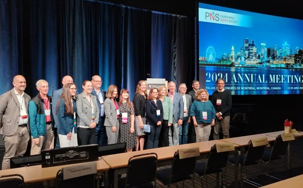
During the 2024 Peripheral Nerve Society meeting (pnsociety.com), held in Montreal (Canada), one of ENU…

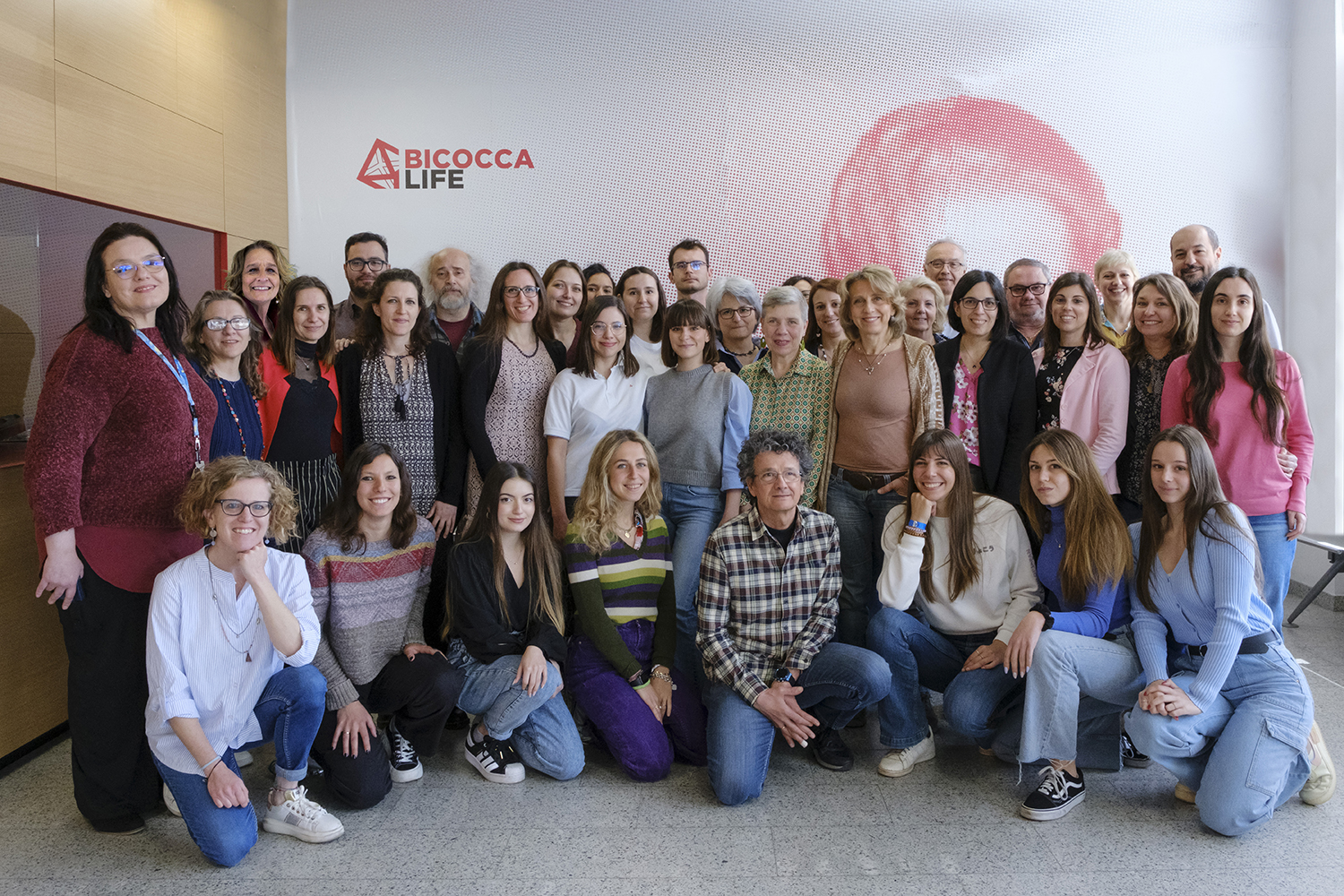
Experimental Neurology Unit
During the 2024 Peripheral Nerve Society meeting (pnsociety.com), held in Montreal (Canada), one of ENU…
One of ENU members, Paola Alberti, MD, PhD, is taking part in the Multinational Association…
The 2024 annual PNS (Peripheral Nerve Society) meeting will take place in Montreal (Canada) from…
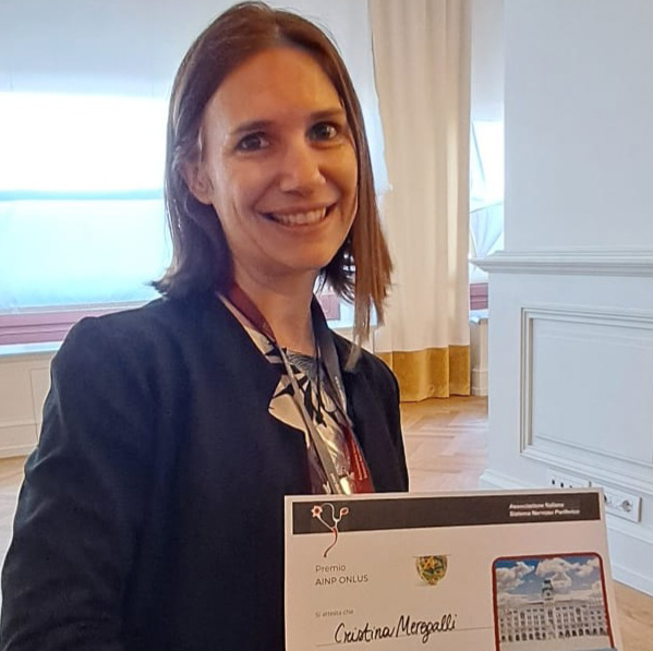
During the Fourteenth Annual Meeting of the Italian Association for the Study of the Peripheral…
The 14th meeting of the ASNP (Italian association for the study of the peripheral nervous…
The National Institute of Health is organising a series of webinar titled Building bridges in…